

Primary Health Insurance:
BlueCross BlueShield
United Health Care
Humana
Cigna
North Mississippi Acclaim
Medicare
Medicaid


If you have recently undergone eye surgery, your eye doctor may recommend the use of post-surgical contact lenses as part of your recovery process. These specialized lenses are designed to aid in the healing and protection of your eyes after surgery.
After eye surgery, your eyes are in a delicate state and require extra care to ensure proper healing. Post-surgical contact lenses can play a crucial role in this process. These lenses act as a protective barrier, shielding your eyes from irritants such as dust, debris, and bright lights. They also help to maintain the shape of the cornea and aid in the prevention of infection. By wearing post-surgical contact lenses, you can minimize the risk of complications and promote a faster and smoother recovery.
There are various types of post-surgical contact lenses available, and the specific type recommended for you will depend on the nature of your surgery and your eye condition. One common type is the bandage contact lens, which is a soft, therapeutic lens that covers the cornea, providing protection and promoting healing.
Another type is scleral lenses, which is larger and covers a larger portion of the eye, providing enhanced protection and support. Your optometrist will determine the most suitable type of post-surgical contact lens for your individual needs.

Contact lenses have become a popular choice for individuals who want to correct their vision without the hassle of wearing glasses. Traditional contact lenses have been around for decades, offering a convenient alternative to eyeglasses. However, advancements in technology have given rise to a new type of contact lens – hybrid contacts.
Hybrid contacts are a revolutionary type of contact lens that combine the best features of both soft and rigid gas permeable (RGP) lenses. The rigid center corrects vision by providing precise clarity, while the soft skirt offers comfort and stability. This unique combination allows for the benefits of both types of lenses to be experienced simultaneously.
The central RGP lens of a hybrid contact is made from a rigid material that allows oxygen to pass through to the cornea, ensuring ample oxygen supply to the eyes. This ensures the overall health of the eyes, preventing dryness and reducing the risk of complications associated with limited oxygen flow.

Post-concussion syndrome (PCS) is a complex condition that can occur after a traumatic brain injury (TBI), such as a concussion. While the initial injury may have healed, the lingering symptoms can significantly impact your daily life, making it challenging to return to your normal routines and activities.
Post-concussion syndrome is a collection of physical, cognitive, and emotional symptoms that can persist for weeks, months, or even years after a head injury. Unlike the typical recovery from a concussion, which usually resolves within a few days or weeks, PCS can be a prolonged and debilitating experience. The symptoms of PCS can vary widely from person to person, making it a complex and often misunderstood condition.
One of the primary causes of post-concussion syndrome is the disruption of the brain’s normal function due to the injury itself. When the brain experiences trauma, it can lead to a variety of biochemical and structural changes, which may cause the ongoing symptoms of PCS. The severity of the concussion does not always correlate with the likelihood of developing PCS; even minor concussions can lead to prolonged post-concussion symptoms in some individuals.
Another contributing factor to PCS may be related to the individual’s psychological response to the injury. Anxiety, stress, and pre-existing mental health conditions can exacerbate the symptoms of PCS and prolong the recovery process. People who have a history of migraines, anxiety disorders, or depression may be more prone to developing post-concussion syndrome.
The symptoms of post-concussion syndrome can be diverse and can vary in severity from person to person. Some of the most common symptoms include:
Physical Symptoms: Headaches, dizziness, nausea, sensitivity to light or sound, fatigue, and sleep disturbances.
Cognitive Symptoms: Difficulty concentrating, memory problems, slower processing speed, and reduced problem-solving abilities.
Emotional Symptoms: Irritability, mood swings, anxiety, depression, and increased emotional sensitivity.
The presence and severity of these symptoms can fluctuate over time, making it crucial to monitor your condition closely and communicate any changes to your primary care or eye doctor.

Cataracts are a common eye condition that can significantly affect your vision. They occur when the lens of your eye becomes cloudy, causing blurred or distorted vision. This clouding is typically a result of aging, but other factors such as genetics, diabetes, or prolonged exposure to sunlight can also contribute to the development of cataracts.
As cataracts progress, you may experience symptoms such as difficulty seeing at night, increased sensitivity to glare, or a decrease in color perception. These changes can make daily activities like reading, driving, or recognizing faces more challenging.
Cataract surgery is often recommended when cataracts start to interfere with your vision and affect your ability to perform daily tasks. The primary goal of cataract surgery is to remove the cloudy lens and replace it with an artificial intraocular lens (IOL).
The decision to undergo cataract surgery is a personal one and should be made after consulting with your optometrist. Factors such as the severity of your cataracts, your overall eye health, and your lifestyle requirements will be taken into consideration when determining if surgery is necessary.
Cataract surgery co-management refers to the collaborative approach between an ophthalmologist, who performs the surgery, and an optometrist, who provides pre-operative and post-operative care. This partnership ensures that you receive comprehensive eye care throughout the entire process, from the initial consultation to the final follow-up appointments.
Your optometrist plays a crucial role in cataract surgery co-management by conducting a thorough pre-operative evaluation to assess your overall eye health and determine the most suitable IOL for your specific needs. They will also provide guidance and education on the surgical procedure, address any concerns you may have, and help you make informed decisions about your eye care.
Technology continues to blur the lines between style and functionality - and Nuance Audio Glasses are a perfect example of this evolution. Designed to look like everyday eyewear, these innovative glasses feature built-in hearing enhancement technology that helps users hear conversations clearly while maintaining natural, effortless vision.
Nuance Audio Glasses are smart eyeglasses designed to provide both vision correction and enhanced hearing. Developed by Nuance Hearing, these glasses discreetly integrate directional microphones, miniature speakers, and Bluetooth connectivity into the frame - allowing wearers to improve their hearing experience without traditional hearing aids. By combining hearing technology with fashionable eyewear, Nuance Audio Glasses offer a seamless, everyday solution for individuals who want better hearing support without drawing attention to their device.
The technology behind Nuance Audio Glasses focuses on selective sound amplification and speech clarity. The glasses use beamforming microphones to focus on voices in front of the wearer while reducing background noise. The enhanced sound is then transmitted through tiny speakers near the ears, allowing users to hear clearly without the occlusion effect common with in-ear devices.
This open-ear design keeps the ear canals unobstructed, promoting natural hearing while boosting important speech frequencies - ideal for conversations in noisy environments like restaurants or social gatherings.

Newton™, formerly Neurolens®, is the first and only prescription lenses that include an element of contoured prism in their design. This prism is designed to bring the patient’s eyes into more equal alignment, and this should help to provide relief from the symptoms that are associated with several eye misalignment conditions, including digital eye strain and binocular vision dysfunction.
What is digital eye strain?
Digital eye strain is the name given to describe a group of symptoms that can occur when someone spends long periods of time using digital devices. Since using digital devices requires the eyes to work harder than normal and we don’t always position our devices the perfect distance away, it can lead to issues such as eye pain, dry and irritated eyes, eye fatigue, light sensitivity, and blurred vision. Unsurprisingly, the number of people who are experiencing digital eye strain has grown significantly over the last few years and is expected to continue to do so.
What is binocular vision dysfunction?
Binocular vision dysfunction, also known as BVD for short, is another eye condition but is one that is very misunderstood. Binocular vision dysfunction occurs when the eyes aren’t perfectly aligned, causing your brain and eyes to work harder than normal in order to create a clear visual image and remain focused. This places pressure on the trigeminal nerve, which is the nerve that is responsible for the majority of the sensations that we experience in our head and back. BVD can often manifest as other things owing to the huge range of symptoms that are associated with the condition. These can include, but aren’t limited to:
Blurred vision
Headaches/migraines
Double vision
Motion sickness
Vertigo
Dizziness
Anxiety
Many people don’t think to visit an eye doctor when they are experiencing these symptoms, but all can occur simply because the eyes are out of alignment.

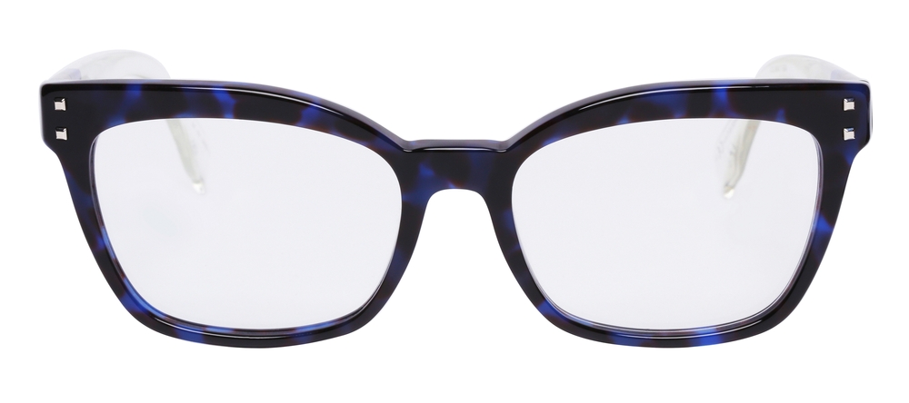
Colored contacts have become increasingly popular over the years as a way to enhance your look and experiment with different eye colors. Whether you want to change your eye color for a special occasion or simply for everyday wear, colored contacts offer a fun and versatile option.
When it comes to colored contacts, there are various types to choose from, depending on your preferences and needs. The three main categories are cosmetic, enhancement, and opaque lenses.
Cosmetic lenses are designed to completely change the color of your eyes. These lenses come in a wide range of shades, from natural hues such as blue, green, and brown, to more vibrant colors like purple, gray, and even red. Cosmetic lenses can create a dramatic transformation and give you the eye color you desire.
Enhancement lenses, on the other hand, are designed to enhance your natural eye color rather than change it completely. They add depth and intensity to your eyes, making them appear brighter and more vibrant. Enhancement lenses are ideal if you want a subtle change that enhances your natural beauty.
Opaque lenses are specifically designed to cover dark-colored eyes. They are ideal for individuals with naturally dark eyes who want to achieve a lighter or more vibrant eye color. Opaque lenses are often used in theatrical or costume applications as they can create a striking and captivating look.

Telemedicine makes it easier to connect with your optometrist when an in-office visit is not necessary or not possible. A virtual eye care appointment allows you to discuss certain eye health concerns, follow-up needs, and symptoms through a secure video visit from home, work, or another private location.
All you need is a smartphone, tablet, or computer with a camera, microphone, and internet connection. The team will provide instructions before your appointment so you know what to expect and how to join your virtual visit.
To schedule a telemedicine appointment, call our office or request an appointment online. Before confirming your visit, we may ask a few questions about your symptoms, medical history, and current eye care concerns. This helps us determine whether a virtual visit is appropriate or if you should be seen in person.
During your appointment, your optometrist will review your concerns, ask about your symptoms, and may guide you through simple observations or questions to better understand what is happening. In some cases, photos of the eye or eyelid may be helpful if they are clear and taken safely.

Technology continues to blur the lines between style and functionality - and Nuance Audio frames are a perfect example of this evolution. Designed to look like everyday eyewear, these innovative glasses feature built-in hearing enhancement technology that helps users hear conversations clearly while maintaining natural, effortless vision.
Nuance Audio are smart eyeglasses designed to provide both vision correction and enhanced hearing. Developed by Nuance Hearing, these glasses discreetly integrate directional microphones, miniature speakers, and Bluetooth connectivity into the frame - allowing wearers to improve their hearing experience without traditional hearing aids. By combining hearing technology with fashionable eyewear, Nuance Audio glasses offer a seamless, everyday solution for individuals who want better hearing support without drawing attention to their device.
The technology behind Nuance Audio glasses focuses on selective sound amplification and speech clarity. The glasses use beamforming microphones to focus on voices in front of the wearer while reducing background noise. The enhanced sound is then transmitted through tiny speakers near the ears, allowing users to hear clearly without the occlusion effect common with in-ear devices.
This open-ear design keeps the ear canals unobstructed, promoting natural hearing while boosting important speech frequencies - ideal for conversations in noisy environments like restaurants or social gatherings.
What sets Nuance Audio apart is their easy-to-use ability ability to personalize and fine-tune your hearing experience directly from your smartphone. Through the Nuance Audio app, users can adjust sound levels, change listening modes, and even customize the direction of sound focus. Whether you’re watching TV, attending a meeting, or spending time outdoors, you can easily switch between settings for optimal clarity and comfort - all from the palm of your hand.

We all want to look our best and in the last decade, we have seen a significant increase in the number of people seeking cosmetic services in order to enhance their appearance. With our eyes being our most distinguishing feature, we want to make the most of them. Thankfully there is now a range of cosmetic services that can help to rejuvenate our eyes and the area around them to keep them fresh, young and wrinkle-free.
Let’s take a look at some of the services on offer.
The brown pigment spots that appear on the face are often referred to as age spots and are a result of sun exposure. With age, the repeated exposure to UV rays causes melanin, a compound that is responsible for pigmentation and protecting the skin begins to clump together to form an area of hyperpigmentation. Whilst they aren’t any cause for concern, many people feel that they look unsightly. Luckily, there are a number of different treatments that you can get to remove them including topical creams, laser therapy, and chemical peels.
If you are suffering from darker pigmentation then we strongly recommend that you make an appointment with a qualified dermatologist who will recommend the best course of treatment for you, based on your specific needs.
08:30 AM - 4:30 PM
09:00 AM - 3:00 PM
CLOSED